Diabetic wounds heal slowly due to nerve damage, poor blood flow, and infection risk. Learn why diabetic wounds don’t heal and when to get help.
People with diabetes often notice that even small cuts, blisters, or sores take much longer to heal. Some wounds may not heal at all. Others may worsen quietly and lead to serious complications before pain or warning signs appear.
Diabetic wounds do not heal slowly by accident. Diabetes affects the body in ways that directly interfere with wound healing. High blood sugar damages nerves, weakens circulation, and reduces the body’s ability to fight infection. When these problems overlap, healing becomes difficult.
Understanding why diabetic wounds don’t heal helps patients and caregivers recognize risk early and take action before wounds become severe.
This guide explains how diabetes affects wound healing, the role of nerve damage and circulation problems, why infection risk is higher, and how diabetic wounds can progress if left untreated.
Find a wound care specialist near you
Diabetes and Wound Healing
Normal wound healing depends on several key processes working together. Blood must deliver oxygen and nutrients. Immune cells must control bacteria. New tissue must grow and repair damage.
Diabetes interferes with all of these steps.
High blood sugar levels damage small blood vessels over time. This limits oxygen delivery to the skin. Without oxygen, healing slows.
Diabetes also affects immune function. White blood cells become less effective at fighting bacteria. This makes wounds more likely to become infected and harder to clear once infection begins.
In addition, diabetes disrupts collagen production. Collagen is a protein that helps rebuild skin and tissue. When collagen production is impaired, wounds close more slowly and remain fragile.
These changes mean that wounds in people with diabetes often heal differently than in people without diabetes, even when the wounds look similar at first.
Key Takeaway
Diabetic wounds don’t heal because diabetes affects nerves, circulation, immune response, and tissue repair. These changes allow small injuries to worsen quietly and heal slowly.
Understanding these risks empowers patients to act early. With proper care, many diabetic wounds can be treated before they become severe.
Nerve Damage and Injuries
Neuropathy
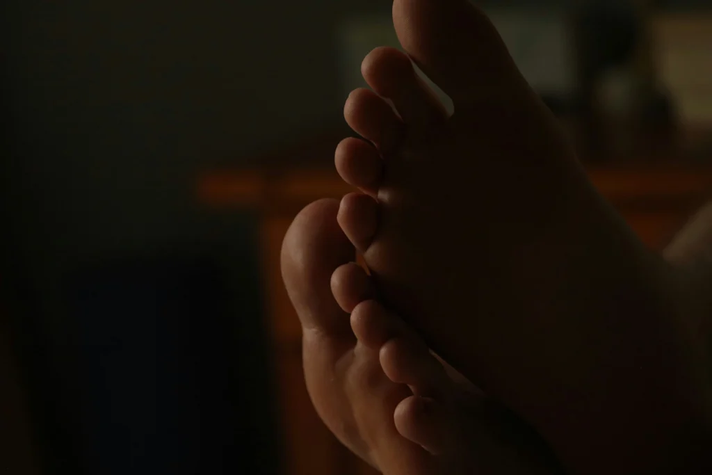
One of the most important reasons diabetic wounds don’t heal is neuropathy, or nerve damage. Diabetic neuropathy reduces sensation, especially in the feet and lower legs. People may not feel pain, pressure, heat, or small injuries. As a result, wounds often go unnoticed.
Common examples include:
- Blisters from shoes
- Cuts from trimming nails
- Cracks in dry skin
- Pressure sores from walking or standing
Without pain as a warning signal, people continue to walk or apply pressure to injured areas.
This repeated stress prevents healing and deepens the wound.
Neuropathy can also affect sweat glands [3]. Dry skin cracks more easily, creating openings for bacteria. Over time, small injuries can turn into chronic wounds.
Circulation Problems
Poor Blood Flow
Good circulation is essential for wound healing. Blood carries oxygen, nutrients, and immune cells needed to repair tissue.
Diabetes damages blood vessels and increases the risk of peripheral artery disease (PAD).
When blood flow is reduced, wounds receive fewer resources to heal.
Poor circulation leads to:
- Slower healing
- Cooler skin temperature
- Pale or bluish skin
- Increased tissue damage
In severe cases, tissue may begin to die due to lack of oxygen. This can cause wounds to stall completely or worsen even with proper care.
Poor blood flow also limits how well antibiotics reach the wound, making infections harder to treat.
Infection Risk in Diabetes
People with diabetes have a higher risk of wound infection for several reasons.
High blood sugar weakens immune defenses. White blood cells respond more slowly and kill bacteria less effectively. This allows infections to grow more easily.
Reduced sensation means wounds may go untreated longer. Moist or open wounds provide an ideal environment for bacteria.
Infected diabetic wounds may:
- Drain fluid or pus
- Develop foul odor
- Become red or swollen
- Heal very slowly
- Spread into deeper tissue
Once infection reaches muscle or bone, treatment becomes much more complex. Early infection control is critical to prevent serious complications.
How High Blood Sugar Affects Healing at Every Stage
High blood sugar does more than raise glucose numbers. It changes how the body responds to injury at every step of healing.
Inflammation Lasts Too Long
Inflammation is the body’s first response to injury. It helps clean the wound and fight bacteria. In diabetes, inflammation often lasts longer than it should.
When inflammation does not shut off:
- Tissue repair is delayed
- Swelling increases
- New skin forms more slowly
Prolonged inflammation keeps wounds stuck in the early phase of healing instead of moving forward.
New Tissue Forms More Slowly
After inflammation, the body builds new tissue. This step depends on healthy cells, oxygen, and protein.
High blood sugar interferes with:
- Cell growth
- Collagen formation
- Blood vessel repair
As a result, wounds may look stable but fail to shrink or close. Healing appears “stalled,” even with good care.
Skin Becomes More Fragile
Over time, diabetes weakens skin structure. Skin becomes thinner and less elastic, especially on the feet and lower legs.
Fragile skin:
- Tears more easily
- Breaks down under pressure
- Heals more slowly once injured
This makes repeat wounds more likely, even after healing.
Why Diabetic Foot Wounds Are Especially Common
The feet are the most common site for diabetic wounds, and this is not random.
Several risk factors overlap in the feet:
- Reduced sensation from neuropathy
- High pressure from walking
- Poor circulation in the lower legs
- Dry skin and callus formation
Small injuries in the feet are easy to miss. Continued walking adds repeated stress, which prevents healing.
Calluses and Pressure Points
Calluses form where pressure is repeated. In diabetes, calluses can hide underlying tissue damage.
Under a callus:
- Pressure builds
- Skin breaks down
- Ulcers may form without warning
Routine foot checks help catch these issues early.
How Diabetic Wounds Progress Over Time
Understanding progression helps patients recognize when a wound is becoming serious.
Early Stage
- Skin may crack, blister, or redden
- Pain may be mild or absent
- Healing appears slow
Intermediate Stage
- Wound deepens
- Drainage may appear
- Surrounding skin becomes fragile
- Advanced Stage
- Ulcer forms
- Infection risk rises
Tissue damage may extend to muscle or bone
The earlier care begins, the easier it is to stop this progression.
Common Warning Signs Patients Often Miss
People with diabetes often overlook early warning signs because they seem minor.
Signs that deserve attention include:
- A sore that does not improve after several days
- Redness that spreads slowly
- Drainage on socks or bandages
- Swelling without pain
- Skin that feels warmer or cooler than normal
Any of these may signal a developing wound.
Ulcer Formation
Diabetic wounds often progress into ulcers, especially on the feet.
A diabetic ulcer is an open sore that forms when pressure, injury, or skin breakdown does not heal properly [6]. Ulcers commonly develop over pressure points such as:
- The ball of the foot
- Heels
- Toes
- Sides of the foot
Ulcers form because neuropathy reduces pain, poor circulation limits healing, and pressure continues without relief.
Once an ulcer forms, healing may take weeks or months. Without treatment, ulcers can deepen and become infected.
Ulcers are one of the leading causes of hospitalization and amputation in people with diabetes.
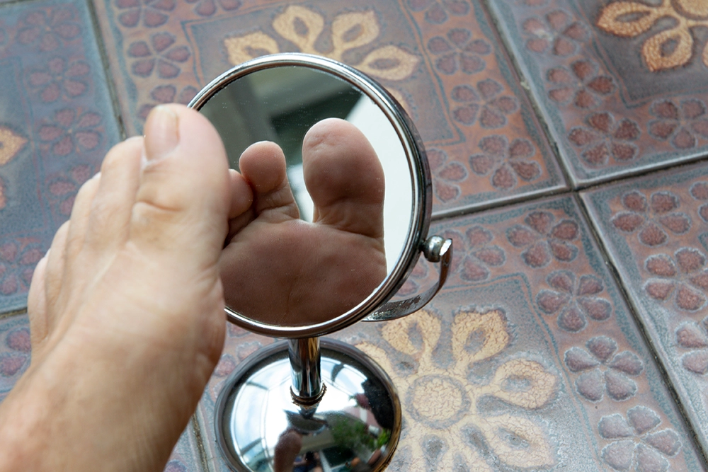
Daily Foot Checks: Why They Matter
Daily foot checks are one of the most effective prevention tools.
Patients should look for:
- Cuts or cracks
- Blisters
- Color changes
- Calluses
- Drainage or odor
Using a mirror or asking for help makes checks easier. Catching changes early prevents major complications.
Why Diabetic Wounds Heal Differently
Healing Factor Without Diabetes With Diabetes Sensation Pain alerts injury Injuries often go unnoticed Blood flow Delivers oxygen efficiently Reduced circulation slows healing
Immune response Controls bacteria Infection risk is higher Tissue repair Collagen forms normally Repair is delayed
Healing speed Predictable timeline Often slow or stalled
Why Waiting Makes Treatment Harder
Many patients delay care because wounds do not hurt or seem small. In diabetes, waiting allows damage to deepen.
Delays can lead to:
- Infection
- Ulcer formation
- Hospitalization
- Surgical treatment
Early evaluation often prevents invasive treatment later.
How Education Reduces Complications
Education is one of the most powerful tools in diabetic wound care.
Patients who understand:
- Why wounds heal slowly
- What changes matter
- When to seek help
…are more likely to act early and avoid severe outcomes.
Why Diabetic Wounds Often Return
Even after healing, risk remains.
Reasons include:
- Ongoing neuropathy
- Persistent circulation problems
- Scar tissue weakness
- Continued pressure points
Prevention does not end when the wound closes. Ongoing care matters.
Why Diabetic Wounds Often Worsen Quietly
One of the most dangerous aspects of diabetic wounds is how quietly they can worsen.
Many people expect pain or redness as warning signs. In diabetes, these signs may be mild or absent.
A wound may:
- Look small on the surface
- Have deep tissue damage underneath
- Show little pain despite worsening
- Progress before visible changes occur
This delayed awareness allows wounds to advance before treatment begins. Regular inspection is essential. Understanding why diabetic wounds don’t heal helps patients prevent ulcers and long-term complications.
The Role of Blood Sugar Control
Blood sugar levels play a direct role in wound healing.
Poor glucose control:
- Slows cell repair
- Increases inflammation
- Feeds bacteria
- Worsens circulation damage
Better blood sugar control improves healing outcomes and lowers infection risk. While control alone cannot heal all wounds, it is a critical part of prevention and treatment.
Why Diabetic Wounds Take Longer to Heal
Even with proper care, diabetic wounds often heal more slowly.
Reasons include:
- Reduced oxygen delivery
- Impaired immune response
- Ongoing pressure or trauma
- Delayed detection
- Existing tissue damage
Healing may require:
- Specialized wound care
- Pressure offloading
- Blood flow evaluation
- Infection management
Understanding that slow healing is common helps patients seek help earlier rather than waiting.
When Diabetic Wounds Become Dangerous
Diabetic wounds become dangerous when:
- They do not improve over time
- Infection develops
- Tissue turns black or necrotic
- Pain suddenly worsens or disappears
- Fever or systemic symptoms appear
In severe cases, infection can spread to bone or bloodstream. Early evaluation reduces the risk of permanent damage.
Why Early Care Makes a Difference
Early care prevents small wounds from becoming major problems.
Simple steps can make a large impact:
- Daily foot checks
- Prompt cleaning and protection
- Pressure relief
- Early medical evaluation
Most serious diabetic wound complications begin as small, manageable injuries.
What Patients Can Do Daily
Daily habits play a key role in preventing non-healing wounds.
Patients should:
- Inspect feet daily
- Keep skin clean and moisturized
- Avoid walking barefoot
- Wear properly fitting shoes
- Manage blood sugar
- Report wounds early
Education and consistency are powerful tools.
When to Get Help
- A wound does not heal within two weeks
- Redness, swelling, or drainage appears
- Pain increases or sensation changes
- Skin turns dark or black
- You have diabetes and any open sore
Waiting too long increases the risk of serious complications.
Concerned About Diabetic Foot Wounds?
If you or someone you care for has diabetes and a wound that isn’t healing, early evaluation can prevent serious complications. Learn how diabetic foot wounds are treated and when medical care is needed.
Find a wound care specialist near you
References:
- Dasari, N., Jiang, A., Skochdopole, A., Chung, J., Reece, E. M., Vorstenbosch, J., & Winocour, S. (2021). Updates in Diabetic Wound Healing, Inflammation, and Scarring. Seminars in plastic surgery, 35(3), 153–158.
- Hammi C, Yeung B. Neuropathy. [Updated 2022 Oct 15]. In: StatPearls [Internet]. Treasure Island (FL): StatPearls Publishing; 2025 Jan
- National Institute of Diabetes and Digestive and Kidney Diseases. (2018). Autonomic neuropathy. U.S. Department of Health and Human Services.
- Ozgok Kangal, M. K., & Kopitnik, N. L. (2025). Physiology, wound healing. In StatPearls. StatPearls Publishing.
- Zhou, K., & Lansang, M. C. (2024). Diabetes mellitus and infection. In K. R. Feingold, R. A. Adler, S. F. Ahmed, et al. (Eds.), Endotext. MDText.com, Inc.
- Wang, X., Yuan, C. X., Xu, B., & Yu, Z. (2022). Diabetic foot ulcers: Classification, risk factors and management. World journal of diabetes, 13(12), 1049–1065.
- Armstrong, D. G., Tan, T. W., Boulton, A. J. M., & Bus, S. A. (2023). Diabetic Foot Ulcers: A Review. JAMA, 330(1), 62–75.
- Berbudi, A., Rahmadika, N., Tjahjadi, A. I., & Ruslami, R. (2020). Type 2 Diabetes and its Impact on the Immune System. Current diabetes reviews, 16(5), 442–449.
 Bleeding is part of the body’s natural response to injury. When skin is cut or damaged, blood vessels constrict and platelets form a clot to seal the wound.
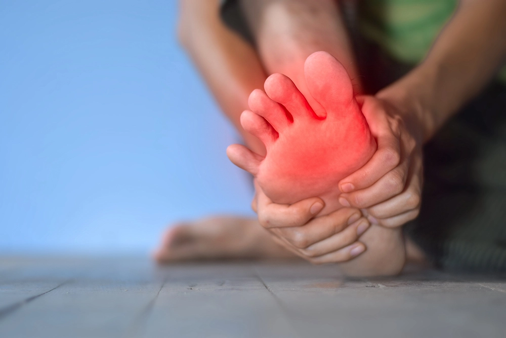 A burning sensation in the feet at night can be uncomfortable, distracting, and even frightening. Many people describe it as tingling, stinging, aching, or feeling like their feet are on fire. For some, the sensation worsens when they lie down, making sleep difficult.