Wound Dock™

Why is your wound bleeding and not stopping? Learn common causes  of persistent wound bleeding, warning signs of complications, and when to seek medical care.

Seeing a wound that won’t stop bleeding can be frightening. Most minor cuts and surgical incisions stop bleeding within minutes. So when bleeding continues longer than expected, it raises concern.  Some bleeding is normal in the early stages of injury. But persistent bleeding, repeated bleeding, or bleeding that restarts after stopping may signal an underlying issue.

Understanding why your wound won’t stop bleeding helps you know when it’s normal—and when it’s not.

What Is Normal Wound Bleeding?

Wound InfectedBleeding is part of the body’s natural response to injury. When skin is cut or damaged, blood vessels constrict and platelets form a clot to seal the wound.

For small cuts, bleeding typically slows within a few minutes with steady pressure. Surgical incisions may have light oozing for 24 to 48 hours.

Normal early bleeding may look like:

  • Bright red blood that slows with pressure
  • Light oozing from stitches
  • Small spotting on dressings
  • Mild bleeding after dressing changes

Bleeding becomes concerning when it does not respond to pressure or continues beyond the expected timeframe.

Common Reasons a Wound Keeps Bleeding

Several factors can interfere with clot formation and wound stability.

Inadequate Pressure

The most common reason bleeding continues is insufficient pressure. A clot needs steady, firm pressure for several minutes to stabilize.  Stopping pressure too soon can restart bleeding.

Disrupted Clot

Clots are fragile early on. Picking at scabs, removing dressings too quickly, or bumping the wound can reopen small vessels.

Infection

Infection increases inflammation and may damage fragile new blood vessels, causing bleeding to restart.

Signs infection may be present include:

  • Increasing redness
  • Warmth
  • Swelling
  • Pus-like drainage
  • Fever

If bleeding is accompanied by these symptoms, evaluation is important.

Medications That Affect Clotting

Certain medications can make bleeding harder to control, including:

  • Blood thinners (anticoagulants)
  • Aspirin
  • Antiplatelet medications
  • Some herbal supplements

These medications do not prevent healing, but they can prolong bleeding.

High Blood Pressure

Elevated blood pressure increases pressure inside blood vessels. This can make clots less stable and cause wounds to bleed longer.

Underlying Bleeding Disorders

Some people have clotting disorders that affect the body’s ability to stop bleeding properly. If wounds frequently bleed excessively, medical evaluation is recommended.

Trauma to the Area

Repeated friction, movement, or tension can reopen small blood vessels.

This is common with:

  • Knee or elbow wounds
  • Abdominal incisions
  • Areas exposed to tight clothing
  • Wounds over joints

When Bleeding After Surgery Is Concerning

Mild spotting on a surgical dressing can be normal. However, bleeding is concerning if you notice:

  • Rapid soaking of bandages
  • Bright red blood that continues flowing
  • Increasing swelling or firmness under the incision
  • Sudden severe pain
  • Blood pooling beneath the skin

Bleeding beneath the skin may form a hematoma. This can create pressure and delay healing.

Contact your surgeon if bleeding increases instead of improving. Persistent bleeding after surgery is not common and should always be evaluated.

What to Do if a Wound Won’t Stop Bleeding

If your wound is bleeding:

  • Apply firm, direct pressure with clean gauze
  • Maintain steady pressure for at least 10–15 minutes
  • Do not repeatedly lift the gauze to check
  • Elevate the area if possible
  • Replace soaked dressings without removing pressure for too long

If bleeding does not slow after sustained pressure, seek urgent care.

Avoid applying powders, sprays, or unapproved substances unless directed by a healthcare provider.

When to Seek Immediate Medical Care

Seek emergency care if you notice:

  • Bleeding that does not stop after 15 minutes of firm pressure
  • Large amounts of bright red blood
  • Spurting blood
  • Signs of shock (dizziness, weakness, pale skin)
  • Rapid swelling or expanding bruising
  • Severe pain
  • Deep wounds exposing muscle or bone

These may indicate significant vessel injury or impaired clotting.

Why Bleeding May Restart After It Stops

Some wounds stop bleeding initially, then begin again hours later.

This can happen because:

  • The clot was disturbed
  • Blood pressure increased
  • Movement stretched the wound
  • Infection weakened the tissue
  • The wound was not fully sealed internally

If bleeding repeatedly restarts, the wound may need reinforcement or medical evaluation.

Can Stress or Anxiety Cause Bleeding?

Stress itself does not directly cause bleeding. However, stress can elevate blood pressure temporarily, which may affect clot stability in some cases.  Managing stress supports overall healing but should not replace medical evaluation for persistent bleeding.

How Blood Sugar Affects Bleeding

In people with diabetes, high blood sugar can impair clot formation and delay healing.

Poor glucose control may increase the risk of infection and fragile blood vessels. Monitoring blood sugar carefully during wound healing improves outcomes. Blood sugar is just one piece of the picture.

Risk Factors for Prolonged Wound Bleeding

Certain factors increase the risk that a wound will bleed longer than expected:

  • Use of blood-thinning medications
  • High blood pressure
  • Liver disease
  • Bleeding disorders
  • Vitamin deficiencies
  • Recent surgery
  • Diabetes
  • Smoking

If you have multiple risk factors, report persistent bleeding promptly.

Signs Bleeding Is Becoming Dangerous Watch for:

  • Continuous bright red bleeding
  • Blood soaking through multiple dressings
  • Lightheadedness
  • Shortness of breath
  • Rapid heartbeat
  • Cool or clammy skin

These signs may indicate significant blood loss.

How Doctors Evaluate Persistent Bleeding

If bleeding does not stop, your provider may assess:

  •  Blood pressure
  • Medication history
  • Clotting function
  • Wound depth
  • Vessel injury
  • Signs of infection
  • Hematoma formation

In some cases, lab tests or imaging may be required.

Treatment may include:

  • Suturing
  • Cauterization
  • Medication adjustments
  • Pressure dressings
  • Drainage of hematoma

Preventing Recurrent Bleeding

To reduce the risk of ongoing bleeding:

  • Follow wound care instructions carefully
  • Avoid heavy lifting if advised
  • Protect the area from friction
  • Monitor blood pressure
  • Take medications exactly as prescribed
  • Inform your provider if you are on blood thinners
  • Avoid picking at scabs

Consistency and protection support clot stability.

Emotional Impact of Persistent Bleeding

Bleeding can trigger anxiety quickly. Blood is visible and dramatic, even when the amount is small.

While persistent bleeding should be taken seriously, many cases resolve with proper pressure and evaluation.

Acting calmly and promptly reduces risk.

Why a Wound May Bleed Under the Skin

Not all bleeding is visible. Sometimes blood collects beneath the surface of the skin. This is called a hematoma.

A hematoma may cause:

  • Swelling that feels firm
  • Deep bruising
  • Increasing pressure or tightness
  • Throbbing pain
  • Skin that appears stretched or shiny

Unlike surface bleeding, a hematoma does not drain easily. Instead, blood pools in the tissue.

Small hematomas may resolve on their own. Larger ones may require drainage to prevent delayed healing or infection.

If you notice swelling that continues to grow or feels tense and painful, contact your provider promptly.

Wound Bleeding and Surgical Drains

After certain surgeries, drains are placed to remove excess blood or fluid. Some drainage is expected.

However, contact your surgeon if you notice:

  • A sudden increase in bright red drainage
  • Clots in the drain tubing
  • Rapid filling of the collection bulb
  • Drain output that increases instead of decreases

Drain output should gradually lessen over time. Increasing output may signal ongoing bleeding.

When Wound Bleeding Is Related to Activity

Sometimes a wound bleeds only after movement.

This can happen because:

  • Activity increases blood pressure
  • Muscles pull against stitches
  • Friction irritates fragile tissue
  • Internal sutures are stressed

If bleeding occurs after exercise, lifting, or stretching, reduce activity and monitor closely.

Follow your provider’s timeline for returning to normal movement. Even if a wound looks healed on the surface, deeper layers may still be fragile.

Wound Bleeding in the First 24 Hours

The first 24 hours after an injury or procedure are the most likely time for bleeding

During this phase:

  • Blood vessels are sealing
  • Clots are forming
  • Tissue is inflamed

Mild oozing during this window can be normal. Bright red, continuous bleeding is not.  Keeping the area elevated and minimizing movement during this early phase helps stabilize clot formation.

Wound Bleeding Days Later

Bleeding that starts several days after injury may be caused by:

  • Infection
  • Suture irritation
  • Scab disruption
  • Increased blood pressure
  • Trauma to the area

Bleeding that occurs after the initial healing period should be evaluated if it is more than mild spotting.

How Infection Makes Wound Bleeding Worse

Infection increases inflammation. Inflamed tissue is fragile and more likely to bleed.

Infected wounds may:

  • Appear red and swollen
  • Feel warm
  • Produce thick drainage
  • Bleed easily when touched

Treating infection often improves bleeding stability.

Special Considerations for People on Blood Thinners

If you take anticoagulants, your provider may adjust medication if bleeding becomes frequent.  Never stop blood thinners without medical guidance. Sudden discontinuation can increase the risk of serious complications.

Instead, contact your provider if:

  • Bleeding episodes are frequent
  • You require prolonged pressure to stop bleeding
  • You develop large bruises
  • Bleeding worsens unexpectedly

Medication adjustments are sometimes necessary.

Wound Bleeding and Nutritional Deficiencies

Certain deficiencies can interfere with clotting and vessel strength.

Low levels of:

  • Vitamin K
  • Vitamin C
  • Iron
  • Protein

All may contribute to fragile tissue or delayed clot stability.  If wounds bleed repeatedly or healing is slow, your provider may recommend lab testing.  Balanced nutrition supports both clotting and tissue repair.

Warning Signs of Significant Blood Loss

While most wound bleeding is minor, severe blood loss can become dangerous.

Watch for:

  • Dizziness
  • Weakness
  • Pale or cool skin
  • Rapid heartbeat
  • Shortness of breath
  • Confusion

These symptoms require emergency care. Do not delay seeking help if you feel faint or unwell alongside ongoing bleeding.

How Long Does It Take for a Clot to Fully Stabilize?

A clot begins forming within minutes. However, full stabilization takes longer.

In the first few hours:

  • The clot is soft and easily disrupted
  • Within 24–48 hours:
  • The clot becomes more secure
  • Tissue begins rebuilding

Even after surface healing appears complete, new blood vessels remain delicate.  Avoid disturbing scabs prematurely. They act as natural protection.  When Bleeding Means the Wound Needs Closure Again Sometimes bleeding indicates the wound edges are separating.

If you notice:

  • The incision widening
  • Bleeding from deeper layers
  • Tissue visible beneath the skin

The wound may need reinforcement.

Prompt evaluation may allow:

  • Additional sutures
  • Sterile adhesive strips
  • Pressure dressings
  • Surgical correction if necessary

Early correction improves healing outcomes.

Protecting a Healing Wound From Re-Bleeding

To prevent recurrent bleeding:

  • Follow lifting restrictions carefully
  • Protect the wound from friction
  • Avoid soaking in baths unless cleared
  • Keep dressings secure
  • Monitor blood pressure
  • Take medications as directed

If bleeding episodes continue despite precautions, further evaluation is necessary. If bleeding continues despite these steps, more specialized care may be needed.

When to Consider a Wound Specialist

If your wound:

  • Bleeds repeatedly
  • Does not stabilize
  • Is complicated by infection
  • Shows delayed healing
  • Is associated with circulation issues

A wound care specialist may be helpful.

Specialists assess clot stability, circulation, tissue health, and risk factors that may not be obvious during routine visits.

Early referral often prevents long-term complications.

The Bottom Line

Most wounds stop bleeding quickly with proper pressure. When bleeding continues, restarts, or increases, it may signal infection, clot disruption, medication effects, or vessel injury.  Persistent bleeding should never be ignored. Early evaluation protects healing and prevents complications.

If your wound is bleeding and won’t stop, do not wait for it to resolve on its own. Apply steady pressure and contact a healthcare provider or locate a wound care clinic if bleeding continues.  Early evaluation can prevent infection, blood loss, and delayed healing. Prompt evaluation can prevent complications and support safe recovery.

References:

Cleveland Clinic. (2024, November 27). Hemostasis: What it is & stages. Cleveland Clinic. Retrieved February 15, 2026

Cleveland Clinic. (2024, April 24). Hemorrhage: What it is, causes, symptoms & treatment. Cleveland Clinic. Retrieved February 15, 2026

Halme, A. L. E., Roshanov, P. S., Tornberg, S. V., Lavikainen, L. I., Devereaux, P. J., Tikkinen, K. A. O., … VISION Investigators. (2024). Timing of major postoperative bleeding among patients undergoing surgery: A cohort study. JAMA Network Open, 7(4), Article e244581.

Mayo Clinic Staff. (2024, October 16). Severe bleeding: First aid. Mayo Clinic. Retrieved February 15, 2026

Subramanian, K., Pravallika, M., & Menon, V. (2018). Evidence for stress-induced bleeding in a patient with von Willebrand factor deficiency. Indian Journal of Psychological Medicine, 40(3), 292–295.

Cleveland Clinic. (2024, November 14). Hematoma: Definition, types & treatment.  Cleveland Clinic. Retrieved February 15, 2026

Kaiser Permanente. (2025, October 1). Surgical drain care: Care instructions. Kaiser Permanente Health Encyclopedia. Retrieved February 15, 2026

Yu, J. C., Shliakhtsitsava, K., Wang, Y. M., Paul, M., Farnaes, L., Wong, V., Kim, J., & Thornburg, C. D. (2019). Hematologic manifestations of nutritional deficiencies: Early recognition is essential to prevent serious complications. Journal of Pediatric Hematology/Oncology, 41(3), e182–e185.

Why is there black tissue around your wound? Learn what black wound edges mean, whether it’s dead tissue, infection, or poor circulation, and when to seek medical care.

Seeing black tissue around the edges of your wound can be alarming. You may wonder if it’s a scab, dried blood, or something more serious.  In many cases, black tissue signals that part of the wound has lost blood supply and the tissue has died. This is called necrosis. While not every dark area is dangerous, black wound edges should never be ignored.

Understanding why black tissue forms around a wound can help you act quickly and safely.

What Does Black Tissue Around a Wound Edges Mean?

Black tissue around a wound is often referred to as eschar. Eschar forms when tissue dies and dries out due to lack of oxygen and blood flow.

Healthy healing tissue is usually pink or red. Black or dark brown tissue around edges of a wound means the cells in that area are no longer alive.

Common descriptions include:

  • Hard, dry, black crust
  • Thick dark covering over the wound
  • Black edges around an incision
  • Dark gray or brown patches that do not bleed

This type of tissue does not heal on its own. It often blocks healthy tissue from growing underneath. That’s why early evaluation matters.

Why Black Tissue Forms Around Edges of Surgical or Chronic Wound

There are several common reasons black tissue may develop.

Poor Blood Flow

Wounds need oxygen-rich blood to heal. When circulation is reduced, tissue can die and turn black.

This is more common in:

  • People with diabetes
  • Peripheral artery disease
  • Smoking history
  • Severe swelling
  • Pressure injuries

Poor circulation is one of the most serious causes of black wound edges.

Wound Infection

Severe infection can damage tissue and reduce blood flow, leading to tissue death.

Signs infection may be involved include:

If black tissue is accompanied by these symptoms, urgent care is needed.

Pressure or Trauma

Continuous pressure, especially over bony areas, can cut off circulation. This is common in bedsores (pressure ulcers).

Prolonged pressure can quickly lead to tissue breakdown and black eschar formation.

Delayed Wound Healing

Chronic wounds that do not heal properly may develop necrotic tissue over time. When tissue does not receive enough oxygen, it begins to break down.

Is Black Tissue Around Wound Edges Always Dangerous?

Not all dark areas are emergencies, but they always require evaluation.

Black tissue may be less urgent if:

  • It is small and dry
  • There are no signs of infection
  • Circulation has already been evaluated
  • You are under medical supervision

However, black tissue can become dangerous if:

  • It spreads
  • The surrounding skin becomes red or swollen
  • Pain increases
  • Drainage develops
  • The area becomes soft or foul-smelling

When in doubt, it is safest to seek medical evaluation.

How Doctors Evaluate Black Tissue Around Wound Edges

If you notice black tissue, your provider may assess:

  • Blood flow to the area
  • Signs of infection
  • Depth of tissue damage
  • Underlying medical conditions
  • Blood sugar levels
  • Pressure or friction factors

In some cases, tests such as ultrasound or vascular studies may be needed to evaluate circulation.

What Is Debridement?

Debridement is the removal of dead tissue from a wound. Dead tissue prevents healthy healing and can trap bacteria.

Types of debridement include:

  • Surgical removal
  • Enzymatic (medicated) debridement
  • Autolytic (moisture-assisted) removal
  • Mechanical cleansing

Your provider will determine the safest method. Debridement is often necessary when black tissue is present.

When to Seek Immediate Medical Care for Black Wound Tissue

Seek urgent care if you notice:

  • Rapid spread of black tissue
  • Severe pain
  • Fever over 100.4°F (38°C)
  • Thick, foul-smelling drainage
  • Red streaks spreading outward
  • Numbness or cold skin near the wound
  • Skin turning gray, purple, or black rapidly

These signs may indicate severe infection or compromised circulation.

Can Black Tissue Be Prevented?

Prevention depends on addressing the root cause.

To reduce risk:

  • Manage blood sugar carefully
  • Stop smoking
  • Avoid prolonged pressure on vulnerable areas
  • Keep wounds clean and properly dressed
  • Elevate swollen limbs
  • Follow wound care instructions consistently

Early treatment of circulation problems significantly reduces risk of necrosis.

When a Wound Specialist Is Needed

You should consider seeing a wound specialist if:

  • Black tissue does not improve
  • The wound has been open for more than two weeks
  • You have diabetes or vascular disease
  • The wound is painful and not healing
  • There are repeated infections

Wound specialists evaluate circulation, infection risk, and tissue health. Early referral can prevent more severe complications, including limb-threatening infections.

The Emotional Impact of Seeing Black Tissue

It’s common to feel anxious when you notice dark tissue around a wound. Many people fear amputation or permanent damage.

While black tissue can be serious, many cases improve with early treatment. Acting quickly often prevents more invasive procedures.

The most important step is not ignoring the change.

What Is the Difference Between Black, Yellow, and White Tissue?

When evaluating a wound, color matters. Not all unhealthy tissue looks the same.

  • Black tissue (eschar) usually means the tissue has died due to loss of blood supply or severe damage.
  • Yellow tissue often indicates slough. Slough is soft, moist, and stringy material made up of dead cells and inflammatory debris. Unlike black eschar, slough may be easier to remove.
  • White or pale tissue may signal poor circulation or maceration (overexposure to moisture). Skin that looks white and soggy around a wound may be breaking down from too much drainage.

These differences help explain why treatment varies. Black tissue often requires more urgent evaluation because it blocks healing and may hide deeper damage.

Can Black Tissue Fall Off on Its Own?

Sometimes dry eschar loosens and separates naturally. However, this does not mean the underlying problem is resolved.

If black tissue falls off, you may notice:

  • A deeper open wound underneath
  • Drainage
  • Red or pink tissue that looks fragile
  • Mild bleeding

It is important not to pick or pull at black tissue. Removing it prematurely can expose deeper structures and increase infection risk.  Your provider will decide whether the tissue should be left in place temporarily or removed through debridement.

When Black Tissue Signals a Circulation Emergency

Black tissue caused by poor blood flow is especially concerning.

If the wound is located on the toes, foot, or lower leg and you notice:

  • Cold skin
  • Pale or bluish discoloration
  • Numbness
  • Severe pain at rest
  • Weak or absent pulses

These may indicate critical limb ischemia, a severe reduction in blood supply.  In this situation, immediate medical evaluation is necessary. Restoring circulation may prevent further tissue loss.

Black Tissue After Surgery

If black tissue appears around a surgical incision, it may be related to:

  • Excess tension on the wound
  • Tight sutures restricting blood flow
  • Swelling causing pressure
  • Infection
  • Compromised circulation

Surgical wounds should gradually improve in color and appearance. A darkening edge, especially if it spreads, should be reported to your surgeon.  Early evaluation may prevent widening of tissue damage.

Black Tissue in Diabetic Wounds

People with diabetes are at higher risk for developing black tissue due to reduced circulation and impaired immune response.  High blood sugar damages small blood vessels over time. This reduces oxygen delivery and slows healing.

If you have diabetes and notice dark discoloration around a wound, do not delay care. Early treatment reduces the risk of deeper infection or amputation.  Managing blood sugar carefully during wound healing improves outcomes.

Does Black Tissue Always Need Debridement?

Not always.

In some cases, dry, stable eschar on the heel may be left in place temporarily if there are no signs of infection and circulation is poor. Removing it too early could expose vulnerable tissue.  However, if there are signs of infection, spreading redness, drainage, or softening tissue, removal is usually necessary.

The decision depends on:

  • Blood flow
  • Infection risk
  • Location of the wound
  • Overall health status

This is why evaluation by a trained clinician is important.

Signs Black Tissue Is Getting Worse

Monitor for changes such as:

  • Expansion of the dark area
  • Increasing redness around the edges
  • Swelling
  • Warmth
  • Softening or foul odor
  • Sudden pain increase

Any of these signs suggest worsening tissue damage or infection.  Photographing the wound every few days can help track progression objectively.

How Long Does It Take for Healthy Tissue to Replace Black Tissue?

After dead tissue is removed and the underlying cause is treated, healthy granulation tissue may begin forming within days to weeks.

Healthy tissue typically appears:

  • Red or deep pink
  • Slightly moist
  • Bumpy or textured
  • Free of foul odor

The speed of improvement depends on:

  • Blood flow
  • Infection control
  • Blood sugar management
  • Nutritional status
  • Wound size

Chronic wounds may take several weeks or months to heal fully.

Risk Factors That Increase Necrosis

Certain conditions make black tissue more likely:

  • Diabetes
  • Peripheral artery disease
  • Smoking
  • Advanced age
  • Obesity
  • Immobility
  • Severe infection
  • Pressure injuries

If you have multiple risk factors, wound changes should be evaluated promptly.

When Black Tissue May Be Part of Normal Healing

In rare cases, very small amounts of dark scabbing at the surface may be part of early healing.  True eschar is usually thick, dry, and firmly attached.

If you are unsure whether you are seeing a scab or necrotic tissue, have it evaluated rather than guessing.

Early clarification prevents complications.

How to Protect the Surrounding Skin

Even if black tissue is being treated, protecting the surrounding skin is critical.

  • Keep the area clean and dry as directed
  • Avoid harsh antiseptics unless prescribed
  • Use recommended dressings
  • Reduce pressure on the area
  • Avoid tight footwear if the wound is on the foot
  • Elevate swollen limbs when possible

Healthy surrounding skin helps the wound recover faster.

The Emotional Response to Dark Wound Changes

Black tissue often triggers fear because it looks severe. Many people immediately worry about amputation.

While necrosis can be serious, early intervention dramatically improves outcomes. Most cases, when treated promptly, do not result in limb loss.  Avoid delaying care due to fear. Evaluation provides clarity and a treatment plan. In some cases, emotional fear is tied to a real circulation concern.

When to Ask for a Vascular Referral

If poor circulation is suspected, your provider may refer you to a vascular specialist.

You may need vascular evaluation if:

  • Pulses are weak
  • The wound is on the lower leg or foot
  • Pain occurs at rest
  • The skin is cold or discolored
  • The wound does not improve after debridement

Improving blood flow can significantly change the healing trajectory.

Questions to Ask Your Doctor

If you notice black tissue, consider asking:

  • Is this necrosis or a scab?
  • Do I need debridement?
  • How is my circulation?
  • Could infection be present?
  • Should I see a wound specialist?
  • What warning signs should I monitor at home?

Clear communication helps you participate actively in your care.

The Bottom Line

Black tissue around the edges of a wound usually signals tissue death due to poor blood flow, infection, or prolonged pressure. While not every case is an emergency, black wound edges require medical evaluation.  Early treatment improves healing outcomes and reduces the risk of serious complications.

If you notice black tissue forming around your wound, do not wait for it to resolve on its own.  Contact your healthcare provider or search for a wound care specialist near you for prompt
evaluation. Early assessment can protect healthy tissue, prevent infection, and support safer healing.

References:

  1. Khalid, N., & Azimpouran, M. (2023, March 6). Necrosis pathology. In StatPearls [Internet]. StatPearls Publishing. Retrieved February 15, 2026
  2. National Library of Medicine. (2025, April 8). Eschar. MedlinePlus Medical Encyclopedia.  Retrieved February 15, 2026
  3. Wernick, B., Nahirniak, P., & Stawicki, S. P. (2025). Impaired wound healing. In StatPearls [Internet]. StatPearls Publishing. Retrieved February 15, 2026
  4. Zaidi, S. R. H., & Sharma, S. (2024). Pressure ulcer. In StatPearls [Internet]. StatPearls Publishing. Retrieved February 15, 2026
  5. Cleveland Clinic. (2025, March 13). Gangrene: Symptoms, causes & treatment.  Cleveland Clinic. Retrieved February 15, 2026
  6. Manna, B. (2023). Wound debridement. In StatPearls [Internet]. StatPearls Publishing.  Retrieved February 15, 2026
  7. Pang, H., Huang, G., Xie, Z., & Zhou, Z. (2024). The role of regulated necrosis in diabetes and its complications. Journal of Molecular Medicine, 102(4), 495–505.

A swollen wound can be part of healing—or a warning sign of infection. Learn why wounds swell, common causes, and when swelling needs medical care.

Swelling around a wound is one of the most common changes people notice during healing. A cut, scrape, or surgical incision may look puffy, tight, or raised, especially in the first few days.  This can be uncomfortable and concerning. Many people wonder why a wound is swollen and whether it’s part of healing or a sign of infection.

In many cases, swelling is part of the body’s normal healing response. But swelling that worsens, spreads, or lasts longer than expected may signal infection, fluid buildup, or circulation problems. Understanding why a wound is swollen—and how swelling should change over time—helps prevent small problems from becoming serious complications.

This guide explains common causes of wound swelling, how infection affects swelling, the role of fluid buildup and circulation, and when swelling becomes dangerous and needs medical care.

Causes of Wound Swelling During Healing

Wound swelling occurs when fluid accumulates in the tissue around an injury. This fluid buildup is often caused by inflammation, which is the body’s natural response to tissue damage.

When skin is injured, blood vessels widen and become more permeable. This allows fluid, immune cells, and proteins to move into the area to support healing. As a result, the tissue may appear swollen, firm, or puffy.

Common causes of wound swelling include:

  • Normal inflammatory response
  • Tissue injury or surgery
  • Ongoing pressure or movement
  • Fluid buildup
  • Poor circulation
  • Infection

Swelling alone does not mean a wound is infected. The key is how swelling changes over time.

Inflammatory Response

Inflammation is the body’s first step in healing. It helps clean the wound and protect against bacteria.

During inflammation:

  • Blood flow increases
  • Fluid moves into the tissue
  • Immune cells gather at the wound site

This process causes swelling, redness, warmth, and sometimes mild pain. In early healing, these changes are expected and often peak within the first few days.

Normal inflammatory swelling should:

  • Stay localized near the wound
  • Gradually improve
  • Decrease as healing progresses

When inflammation does not calm down, swelling may persist or worsen instead of improving.

Tissue Damage and Trauma

The amount of swelling often reflects how much tissue was injured. Larger or deeper wounds disrupt more blood vessels and tissue, leading to more fluid leakage.

Swelling may be more noticeable with:

In these cases, swelling may last longer than with minor wounds. As long as swelling slowly improves and no other warning signs appear, it may still be part of normal healing.

Infection-Related Wound Swelling

Wound Infection is one of the most concerning causes of wound swelling. When bacteria multiply in a wound, the immune system responds aggressively. This increases inflammation and fluid production.

Swelling related to infection often behaves differently than normal healing swelling.

Signs Wound Swelling May Be Caused by Infection

Swelling related to infection may:

  • Increase instead of improve
  • Spread beyond the wound edges
  • Feel firm or tight
  • Be accompanied by redness and heat
  • Occur with drainage or foul odor

Infected wounds often show multiple warning signs at the same time.

Other symptoms may include:

  • Worsening pain or tenderness
  • Fever or chills
  • Delayed healing
  • Skin that appears shiny or stretched

Swelling that continues to worsen after the first few days of healing should always be evaluated.

Why Infection Causes More Wound Swelling

Bacteria trigger a stronger immune response. Blood vessels become even more permeable, allowing more fluid to leak into surrounding tissue.

This creates pressure within the tissue, which:

  • Increases pain
  • Slows blood flow
  • Delays healing
  • Forces fluid outward

As swelling increases, the wound environment becomes less stable and more prone to complications.

Fluid Buildup and Circulation Issues

Swelling is not always caused by infection or inflammation alone. Problems with fluid movement and circulation can also cause wounds to swell.

Edema

Edema refers to fluid accumulation in the tissue. It is especially common in the lower legs, ankles, and feet.

Edema-related swelling may:

  • Develop gradually
  • Feel soft or pitting
  • Worsen later in the day
  • Improve with elevation

Edema increases pressure around a wound, making it harder for fluid to drain and for oxygen to reach the tissue.

Wounds surrounded by edema often:

  • Heal more slowly
  • Leak fluid
  • Appear swollen even without infection

Poor Venous Return

Veins carry blood and fluid back toward the heart. When venous return is impaired, fluid pools in the tissue instead of being reabsorbed.

Poor venous return is common in people with:

  • Chronic venous insufficiency
  • Varicose veins
  • Heart failure
  • Prolonged sitting or standing

In these cases, swelling may worsen when the wound is below heart level and improve with rest or elevation.

Poor venous return makes wound swelling harder to resolve and increases the risk of delayed healing.

Poor Circulation

Arterial circulation delivers oxygen and nutrients needed for healing. When circulation is impaired, swelling may persist even when inflammation is mild.

Poor circulation is more common in:

  • Older adults
  • People with diabetes
  • Peripheral artery disease
  • Smokers

Inadequate blood flow limits the body’s ability to clear excess fluid and repair tissue, allowing swelling to linger.

When Wound Swelling is Dangerous

Not all swelling is an emergency, but certain patterns should never be ignored.

Swelling becomes dangerous when it:

  • Rapidly increases
  • Spreads beyond the wound
  • Causes severe pain or tightness
  • Is accompanied by fever or chills
  • Limits movement or sensation
  • Does not improve over time

These signs suggest that swelling may be caused by infection, deep tissue damage, or compromised circulation.

Compartment Pressure and Tissue Damage

Severe swelling can increase pressure within tissue compartments. When pressure rises too high, it restricts blood flow even further.

This can lead to:

  • Tissue death
  • Nerve damage
  • Severe pain
  • Loss of function

Although uncommon, rapidly worsening swelling should be evaluated urgently.

Swelling and Skin Breakdown

Persistent swelling stretches the skin, making it more fragile. Over time, this can cause:

Managing swelling is essential to protecting both the wound and surrounding skin.

How Swelling Changes During Healing

Understanding how swelling should behave helps distinguish normal healing from complications.

Early Healing

  • Swelling appears within hours or days
  • Peaks early
  • Gradually decreases

Delayed Healing

  • Swelling stays the same
  • Improvement stalls
  • Healing slows

Complicated Healing

  • Swelling worsens
  • New symptoms develop
  • Infection risk increases

Trend matters more than appearance on any single day.

How Movement and Position Affect Swelling

Swelling often changes with activity.

Swelling may increase:

  • After walking or standing
  • Later in the day
  • With prolonged sitting

Swelling may decrease:

  • With rest
  • With elevation
  • Overnight

Temporary swelling that improves with rest is often circulation-related rather than infectious.

Why Swelling Can Appear Without Pain

Pain is not always present with wound swelling.

Reduced pain sensation is common in:

  • Diabetes
  • Neuropathy
  • Spinal cord injury
  • Older adults

In these cases, swelling may be the only visible sign of a problem. Visual checks are especially important when pain is unreliable.

How Wound Care Practices Affect Swelling

Wound care choices influence swelling.

Swelling may worsen when:

  • Dressings are too tight
  • Bandages trap moisture
  • Pressure is not relieved
  • Wounds are repeatedly disturbed

Proper dressing fit and pressure relief help control swelling and support healing.

Why Wound Swelling Can Interfere With Wound Healing

Swelling does more than change how a wound looks. Excess fluid around a wound can directly interfere with the healing process. When tissue becomes swollen, pressure builds within the skin and underlying structures. This pressure makes it harder for oxygen and nutrients to reach the wound.

Oxygen is essential for healing. It supports new tissue growth and helps immune cells fight bacteria. When swelling limits oxygen delivery, healing slows. Even wounds that are otherwise well cared for may struggle to improve if swelling remains uncontrolled.

Swelling can also delay healing by:

  • Reducing blood flow to fragile tissue
  • Making wound edges harder to close
  • Increasing tension on healing skin
  • Causing discomfort that limits movement

Over time, persistent swelling keeps wounds stuck in an inflammatory state instead of allowing them to progress into the rebuilding phase of healing.

How Wound Swelling Affects Wound Drainage

Swelling and drainage often occur together. When fluid builds up in surrounding tissue, it increases pressure inside the wound. This pressure can force fluid outward, leading to ongoing leakage or oozing.

Wounds affected by swelling may:

  • Leak clear or pink fluid
  • Soak dressings more quickly
  • Appear wet despite proper care
  • Develop irritation around the wound edges

Drainage caused by swelling does not always mean infection. However, when swelling and drainage increase together, the wound environment becomes less stable. Managing swelling often reduces excess drainage and helps the wound seal more effectively.

Why Wound Swelling Can Return After An Initial Improvement

Many patients notice swelling improves at first, then comes back days later. This can be confusing and discouraging.

Swelling may return when:

  • Activity level increases
  • The wound is placed below heart level
  • Dressings or footwear add pressure
  • Fluid shifts throughout the day

This does not always mean healing has failed. Swelling often fluctuates during recovery. The key concern is whether swelling continues to worsen or stops improving altogether.

Tracking when swelling increases—such as later in the day or after standing—can help determine whether circulation or pressure is contributing rather than infection.

Why Wound Swelling Persists in Chronic Wounds

Chronic wounds remain in an inflammatory state for long periods. This leads to ongoing swelling that may never fully disappear.

In chronic wounds:

  • Fluid cycles in and out
  • Tissue becomes fragile
  • Swelling fluctuates

Changes in swelling are still meaningful. Sudden increases may signal infection or worsening circulation.

Who Is at Higher Risk for Problematic Wound Swelling

Swelling is more likely to become dangerous in people with:

  • Diabetes
  • Poor circulation
  • Heart disease
  • Kidney disease
  • Limited mobility
  • Older age

In these groups, early evaluation is especially important.

What People Can Do to Monitor Wound Swelling

People can help manage wound swelling by:

  • Checking the wound daily
  • Watching for changes in size or firmness
  • Noting when swelling improves or worsens
  • Elevating the area when appropriate
  • Reporting worsening symptoms early

Tracking patterns over time provides valuable information for care decisions.

When to Seek Medical Care

Seek medical care if wound swelling:

  • Increases instead of improving
  • Becomes painful or tight
  • Is accompanied by redness, heat, or drainage
  • Causes fever or chills
  • Interferes with movement
  • Does not improve with basic care

Early care prevents complications and supports healing.

The Role of Elevation in Reducing Wound Swelling

Elevation helps reduce swelling by allowing gravity to assist fluid return toward the heart. This is especially helpful for wounds on the legs, ankles, or feet.

Elevation works best when:

  • The wound is raised above heart level
  • The position is maintained for extended periods
  • It is combined with movement when appropriate

Elevation does not replace medical care, but it can significantly reduce swelling related to fluid pooling. When swelling improves with elevation but returns when the limb is lowered, circulation issues are often involved.

Why Wound Swelling Deserves Early Attention

Swelling is often one of the first visible signs that a wound is under stress. Addressing swelling early helps prevent a cascade of complications.

Early attention to swelling can:

  • Improve oxygen delivery
  • Reduce drainage and skin breakdown
  • Lower infection risk
  • Shorten healing time

Ignoring swelling allows pressure and inflammation to build. Over time, this increases the risk of delayed healing, wound enlargement, and infection.

Key Takeaway

Swelling around a wound is common, especially early in healing, but it should always be monitored closely. Swelling that gradually improves is often part of the body’s repair process.  When swelling spreads, worsens, or begins to limit comfort or movement, it may signal that healing is under strain.

The most important clue is change over time. Swelling that settles as days pass is reassuring, while swelling that lingers or intensifies may point to infection, fluid buildup, or circulation problems. Paying attention to these patterns—and seeking evaluation early—can prevent a manageable wound from becoming more complicated.

Concerned About Infection?

If your wound is swollen and not improving—or if swelling is getting worse—it may be a sign of infection. Learn the early warning signs and when treatment matters.

References:

1. Schilrreff, P., et al. (2022). Chronic inflammation in non-healing skin wounds and advanced therapeutic approaches.[Journal/Publisher]. PMID: 35563319.

2. Ding, X., Tang, Q., Xu, Z., Xu, Y., Zhang, H., Zheng, D., Wang, S., Tan, Q., Maitz, J., Maitz, P. K., Yin, S., Wang, Y., & Chen, J. (2022). Challenges and innovations in treating chronic and acute wound infections: from basic science to clinical practice. Burns & trauma, 10, tkac014.

3. Lent-Schochet, D., & Jialal, I. (2025). Physiology, edema (Updated 2023 May 1). In  StatPearls. StatPearls Publishing.

4. Castera, M. R., & Borhade, M. B. (2025). Fluid management (Updated 2025 April 29). In StatPearls. StatPearls Publishing.

5. Zabaglo, M., Leslie, S. W., & Sharman, T. (2025). Postoperative wound infections (Updated 2024 March 5). In StatPearls. StatPearls Publishing.

In this blog, we will discuss pressure ulcers, and although some may not first recognize the words “pressure ulcer”, most of us know the term “bedsore”, and that is exactly what a pressure ulcer is. Identifying risk factors and knowing what to do about them can prevent problems further down the road.

Overview of Pressure Ulcers

Bedsores
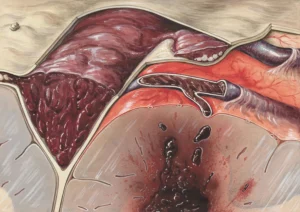
Pressure ulcers, commonly referred to as bedsores, are injuries to the skin and the underlying tissue caused by prolonged pressure on the skin. These wounds typically develop over bony areas of the body, such as the heels, hips, and tailbone. We may also hear them described as pressure ulcers or decubitus ulcers. The formation of bedsores primarily occurs when pressure limits blood flow to the skin, ultimately leading to tissue damage.

While we may typically think of these as issues primarily affecting the elderly or those who are bedridden, pressure ulcers can occur in individuals of any age who experience limited mobility or sensory impairment. The COVID-19 pandemic has significantly impacted the prevalence of pressure ulcers, as many individuals requiring long-term care or hospitalization now experience reduced mobility and increased pressure on their tissues.

Additionally, pressure ulcers have been linked to environmental factors, such as hospital settings where patients may be immobilized for extended periods. During the pandemic, healthcare systems faced unprecedented challenges, and the incidence of pressure ulcers among patients increased dramatically. This not only poses serious health risks but also places an economic burden on the healthcare system, necessitating effective strategies for management and prevention.

Recognizing where bedsores tend to develop can assist in prevention. The most common sites for these ulcers include the back and sides of the head, shoulders, elbows, hips, lower back, and areas around the knees and heels. Various demographic and health factors increase the risk of developing pressure ulcers. Individuals with limited mobility, chronic medical conditions, or those who are bedridden are especially vulnerable. Conditions such as diabetes, vascular disease, and the natural decline in skin resilience associated with aging can also contribute to an increased risk.

Key Causes and Risk Factors

The causation of pressure ulcers consist of a multitude of factors, involving a combination of external and internal forces that contribute to the injury’s formation. Externally, prolonged pressure on the skin leads to decreased blood flow, resulting in tissue damage and ulceration.  Friction from the movement of skin against other surfaces and shear forces, which occur when the skin moves in a different direction than the underlying bone, are also critical factors. A common example of this is patients or elderly who are bedridden for long periods of time, hence the term “bedsores”.

Internal factors such as poor nutritional status, anemia, and chronic diseases can exacerbate the risk for developing pressure ulcers. For example, individuals with diabetes or vascular diseases may suffer from impaired blood flow, reducing the oxygen available to tissues. Age is also a significant risk factor; older adults are more susceptible due to factors like thinner skin and decreased elasticity. Other common risk factors include immobility due to neurological conditions, cognitive issues that impair a person’s ability to reposition themselves, and incontinence, which can lead to skin integrity loss.

Identifying Early Signs

Identifying the early signs of bedsores is crucial for effective intervention and management. Key symptoms to look for include changes in skin color, texture, and temperature. Initially, a bedsore might present as a localized area of skin that appears discolored. On light skin, this may manifest as a red area that does not fade when pressure is applied. In individuals with darker skin, the
discoloration may appear as purple or blue patches.

Aside from changes in color, we must observe texture changes as well. An affected area might feel warm, spongy, or hard to the touch—indications of the skin’s deterioration. We should also pay attention to temperature variation; an area that feels either cooler or warmer than surrounding skin may be an early sign of developing damage. Regularly inspecting the skin can help us identify these symptoms before they progress to more severe stages of pressure ulcers.

Understanding the Staging System

Understanding the staging system for pressure ulcers is vital, as it helps determine the severity of the wounds and informs treatment strategies. The term ‘blanch’ used below means ‘to turn white’. Generally, pressure ulcers are categorized into four stages based on depth and severity.

  • Stage I: The skin is intact but may show non-blanchable redness over a localized area.  The skin can feel different—warmer, cooler, softer, or firmer—than the surrounding tissue.
  • Stage II: This stage involves partial thickness loss of skin, manifesting as a shallow open ulcer with a pink or red wound bed. Blisters can also be present.
  • Stage III: Here, the ulcer extends into the subcutaneous fat layer. It may present as a deep crater, and the drainage may become more significant.
  • Stage IV: The most severe stage, where the ulcer is full thickness and extends into muscle, tendon, or bone. The risk of systemic infection and other complications is significantly heightened at this stage.

Understanding these stages helps us determine appropriate treatment options, making it crucial for caregivers and health professionals to recognize the differences and act promptly to prevent progression.

Complications of Untreated Ulcers

The implications of untreated pressure ulcers extend beyond the immediate discomfort. Pressure ulcers can lead to serious complications, including potentially life-threatening infections.  Cellulitis, a bacterial skin infection, is one such risk; if not addressed, it can progress to more severe infections like osteomyelitis, affecting the bones, or sepsis, a systemic response to infection that can result in organ failure.

Prolonged exposure to untreated pressure ulcers can result in long-term health issues that may be difficult to manage. The consequences could include persistent pain, chronic inflammation, and disability stemming from musculoskeletal complications.

Moreover, the consequences are not only physical; we must also acknowledge the psychological effects of living with pressure ulcers. The ongoing discomfort or disfigurement can lead to anxiety, depression, and a diminished quality of life for affected individuals.

Effective Prevention Strategies

Preventing pressure ulcers requires a proactive and comprehensive approach. One of the most effective strategies is repositioning regularly. Every two hours is a recommended guideline for those who are immobile; careful changes in position can relieve pressure and improve blood circulation.

In addition to repositioning, we must consider nutritional factors as critical components of ulcer prevention. Adequate hydration and a balanced diet rich in proteins, vitamins, and minerals are essential in maintaining skin integrity and overall health. Recognizing the need for balanced nutrition can be especially vital for individuals at higher risk.

Using specialized equipment, such as pressure-relieving mattresses and cushions, can also be beneficial. These products help redistribute weight and reduce direct pressure on vulnerable areas.   Employing these preventive measures can significantly decrease the incidence of pressure ulcers in at-risk populations.

Treatment Approaches

When it comes to treating pressure ulcers, proper wound care is of paramount importance.  General hygiene practices such as keeping the wound clean and dry are essential. This includes gentle cleaning and changing dressings regularly to foster a clean environment that promotes healing.

Medical dressings play a significant role in protecting bedsores and supporting the healing process. Various types of dressings are available, each designed to provide moisture, absorb exudate, and protect the area from infection. Creams and ointments can also be beneficial, especially those with hydrating or barrier properties.

In severe cases, surgical intervention may be necessary. Options may include debridement to remove dead tissue, reconstruction procedures to close extensive wounds, or skin grafts to repair the damaged areas. Choosing the right treatment approach depends on the bedsores’ stage and the overall health of the individual.

Preventive Measures and Best Practices

Preventing pressure ulcers is a critical aspect of patient care. We should all adopt specific routines to maintain skin integrity and prevent the formation of ulcers:

  • Regular Position Changes: For individuals who are bedridden, repositioning every two hours is essential to relieve pressure. Those in wheelchairs should shift their position frequently, ideally every 15 minutes.
  • Skin Care: Keeping the skin clean and moisturized is vital. Routine inspections for any signs of redness or breakdown should be undertaken to catch issues early.
  • Nutrition and Hydration: A balanced diet rich in protein, vitamins, and minerals can support skin health and promote healing. Staying hydrated is equally important.
  • Specialized Equipment: Utilizing pressure-relieving mattresses and cushions can help distribute weight evenly and reduce pressure points, particularly in high-risk patients.
  • Education: Educating caregivers and patients about the importance of pressure ulcer prevention can help ensure everyone is vigilant with care practices.

By implementing these best practices, we can significantly reduce the incidence of pressure ulcers and enhance the quality of care for at-risk individuals.

In summary, recognizing the importance of pressure ulcer management is crucial for improving health outcomes. Through awareness of early signs, understanding staging, and implementing effective prevention and treatment strategies, we can mitigate the impact of these significant health concerns. For healthcare providers and caregivers, it is a crucial responsibility to remain vigilant and proactive in the fight against bedsores, ensuring that individuals, especially those with limited mobility, receive the care they deserve. For patients, understanding the nature and causes of bedsores can prevent future issues before they begin.

Reference

  1. Bedsores (pressure ulcers) – Symptoms and causes – Mayo Clinic
  2. Pressure ulcers (pressure sores) – NHS
  3. Pressure Sores: Treatment, Stages, and Symptoms – WebMD
  4. Bedsores (Pressure Ulcers): Symptoms, Staging & Treatment
  5. Pressure Sores | Bedsores | Pressure Ulcers – MedlinePlus
  6. Pressure Ulcer – StatPearls – NCBI Bookshelf
  7. Pressure Ulcers and Wounds/Injury Management
  8. Sepsis and Pressure Ulcers (Pressure Injuries) – Sepsis Alliance
Are you a wound care provider? Start Here